
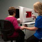
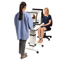
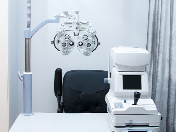
Our Technology
At Colony Eye Care Center our number one priority is the health and well-being of our patients. We have invested in a number of different high-tech imaging and diagnostic devices to ensure our patient have the best and most convenient care.
AdaptDX
This short 3-6 minute test allows our doctors to identify dark adaption – the earliest warning sign of age-related macular degeneration (AMD). AMD impacts daily tasks like: walking; driving; reading; watching tv; recognizing faces; eye-hand coordination (such as putting a key in a door).
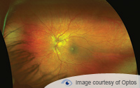
OptoMap Digital Imaging
The OptoMap is an ultra-wide field digital imaging device that allows images of up to 200 degrees of the retina to be taken without dilation. It allows our doctors to make an evaluation of and monitor changes in the retina from year to year. The OptoMap is not a replacement for dilation in the instance of retinal pathology but a very useful tool to aid in diagnosis and increase convenience for the average patient.
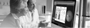
Optical Coherence Tomography (OCT)
Our OCT uses light to capture micrometer-resolution (that’s one-thousandth of a millimeter!), three-dimensional images of the inside of your eyes. The OCT has changed the way we are able to practice and is useful in diagnosing and monitoring macular degeneration, glaucoma, diabetic retinopathy and other retinal pathologies.
Visual Field Analyzer
The visual field is one of the oldest and best tools we have at our disposal. A visual field provides data as to what the patient is actually seeing. It is vital in the management of glaucoma, but can also be useful in monitoring and diagnosing other diseases like retinitis pigmentosa and even brain tumors.
Diopsys Electroretinographer (ERG)
Diopsys machine allows our doctors to evaluate the efficiency of the communication between your eyes and your brain through your optic nerve. It is useful in monitoring glaucoma and convenient for patient’s as it requires no input on their part!
Corneal Topography
The cornea is the front surface of the eye where a contact lens sits. The corneal topographer takes a map of the different curvatures on this surface. Information from this scan helps us in the fitting of contact lenses, especially in “hard” gas permeable, scleral, and corneal refractive tomography lenses. A diagnosis of the disease keratoconus is also made or confirmed with this scan.

© 2026 Colony Eye Care Center. All Rights Reserved. Accessibility Statement | Privacy Policy | Sitemap
Powered by: